Michael Dyer Lab
Examining the coordination of proliferation and differentiation during development and disease
About the Dyer Lab
In the Dyer lab, our fascination focuses on the dynamic molecular and cellular changes that occur during development. We use our deep understanding of normal developmental processes to gain insights into developmental cancers of childhood. The unique opportunity to bridge developmental biology and cancer biology at St. Jude fundamentally changes the way we think of both processes and allows us to forge links between basic biology and medicine. Our research directly contributes to new clinical trials for children with cancer.
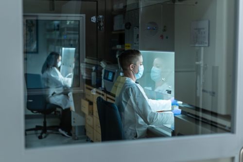
Our research summary
As we explore the bridge between developmental and cancer biology in our lab, we concentrate our efforts on two major areas of research—retinal development and disease and improving outcomes for children with solid tumors.
Retinal development and disease
As progenitor cells exit the cell cycle and differentiate, thousands of genes turn on and off in precisely orchestrated programs. Changes in chromatin structure and genome accessibility accompany those changes in gene expression. Many in the field studied chromatin modifications in embryonic stem cells or cancer cell lines because they are tractable and scalable experimental systems with extensive reference databases. Instead of pursuing this established area of study, we chose to focus on patient tumors and normal-developing human and murine retinae.
The unique research environment at St. Jude allows us to invest the time and effort to overcome the technical hurdles required to develop a map of the chromatin landscape during retinogenesis and in childhood cancers such as retinoblastoma. That investment now yields important new insights into the biology of retinal development and disease.
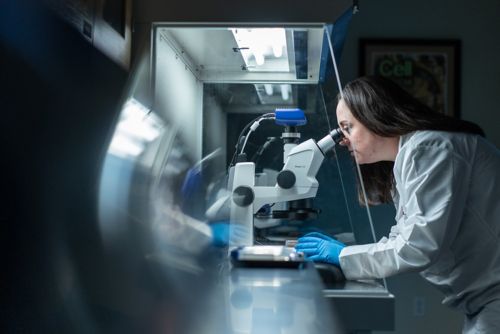
As retinal cells differentiate, they must maintain a group of genes in a state of readiness to turn on at a moment’s notice in response to stress, injury or disease. In mammals, retinal neurons do not regenerate, so they must maintain this state of readiness over their lifetime. The remarkable feature of those rapid-response genes is that they are not normally expressed during retinal development, nor are they required for cellular homeostasis. In normal healthy individuals, they may never activate.
However, if the neurons encounter changes in their environment, toxins, or disease-related stressors, they rapidly activate those transcriptional programs. The particular way that individual cells organize their chromatin during development to respond to stress, injury, or disease later in life is a concept we call cellular pliancy. Looking ahead, we are excited about the new opportunities to manipulate the chromatin landscape in order to alter cellular pliancy in vivo and to visualize the changes in response of individual neurons to stress, injury, or disease.
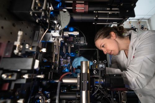
Solid tumor translational research
Our studies on retinal development and disease give us a deep appreciation of the interplay between normal developmental processes and childhood cancer. We extend our studies to include diverse pediatric solid tumors, such as neuroblastoma, osteosarcoma, rhabdomyosarcoma, Ewing sarcoma, and others. Recently, we discovered individual tumor cells can transition through their normal developmental hierarchy, but the process attenuates. These developmental transitions are important because they contribute to disease recurrence after treatment.
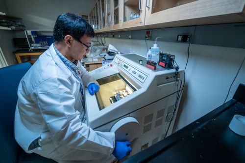
Our current efforts focus on gaining a deeper understanding of the molecular and cellular mechanisms of clonal selection with treatment, with an emphasis on developmental pathways. When patients first receive a diagnosis, we work to eliminate all tumor clones and prevent recurrence by implementing total clonal therapy. We believe this approach will save lives and improve the quality of life for children with solid tumors.
In all our efforts, our lab strives to connect the science to the patient. To achieve this, our diverse and inclusive team of experts guide our multidisciplinary approach. Our diversity drives discovery at every level, and we take pride in this strategy.
Publications
About Michael Dyer
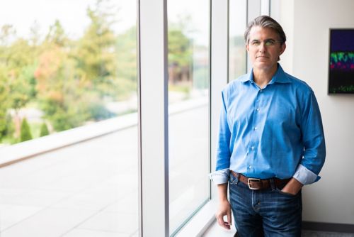
Dr. Michael Dyer draws on his expansive career to guide research in developmental neurobiology. Dyer received his PhD from Harvard University and completed his postdoctoral fellowship at Harvard Medical School where he became interested in the regulation of retinal progenitor cell proliferation during neurogenesis. In 2002, Dr. Dyer began his career at St. Jude, and his interest in neural progenitor cell proliferation grew to encompass cancer biology, evolutionary biology, and stem cell biology. In his time at St. Jude, Dyer has been named a Pew Scholar, a Cogan Award Recipient, and a Howard Hughes Medical Institute Early Career Scientist. In 2013, he became an investigator of the Howard Hughes Medical Institute and a year later he was named the Richard C. Shadyac Endowed Chair in Pediatric Cancer Research. He serves as Chair of the Developmental Neurobiology department and is a co-leader for the Developmental Biology and Solid Tumor program.
The team
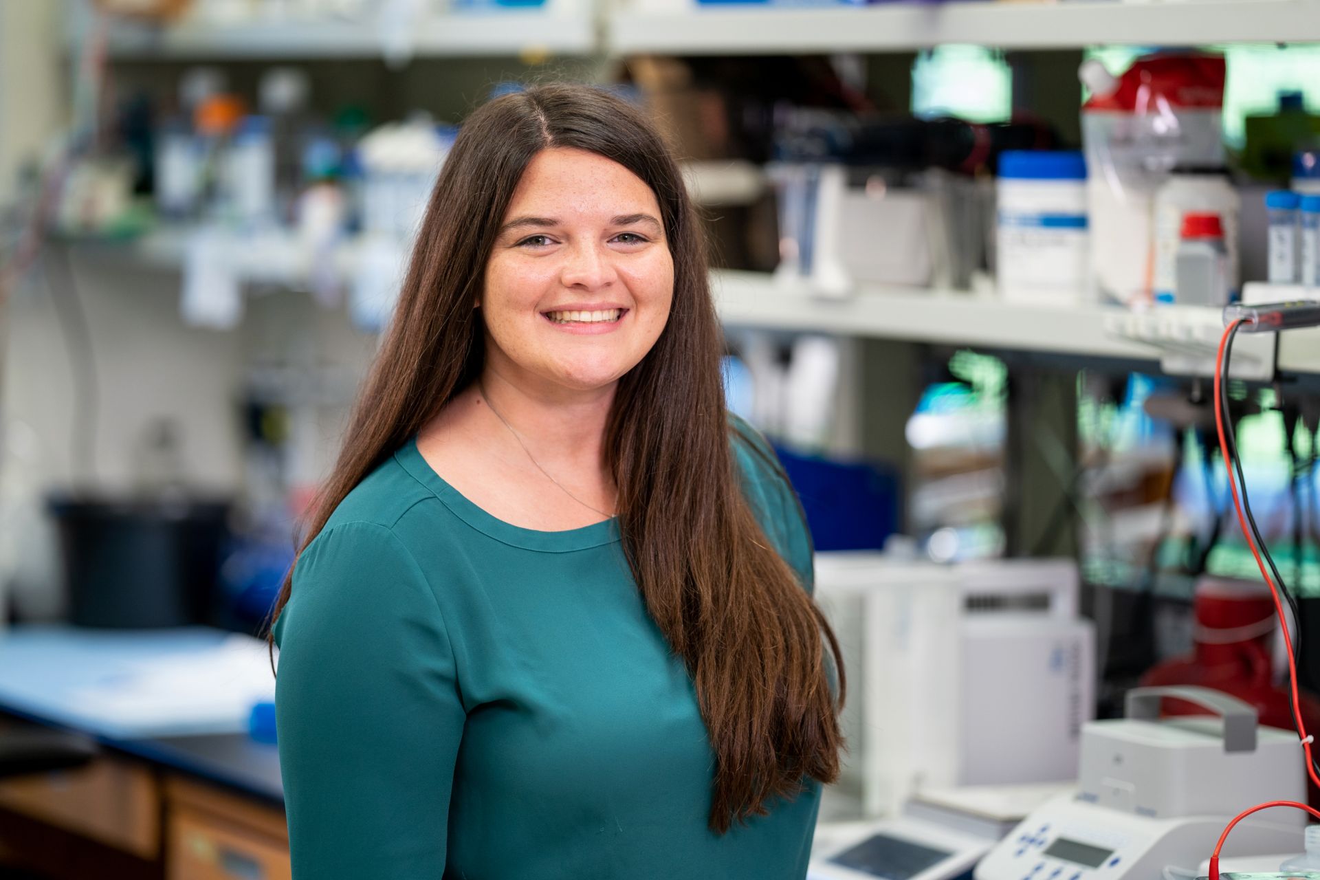
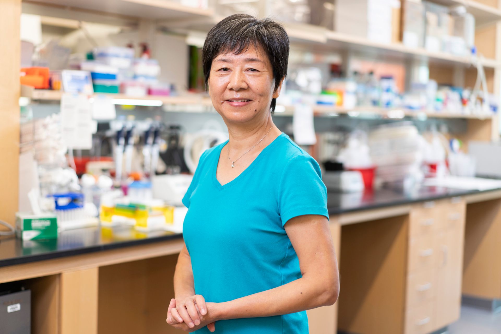
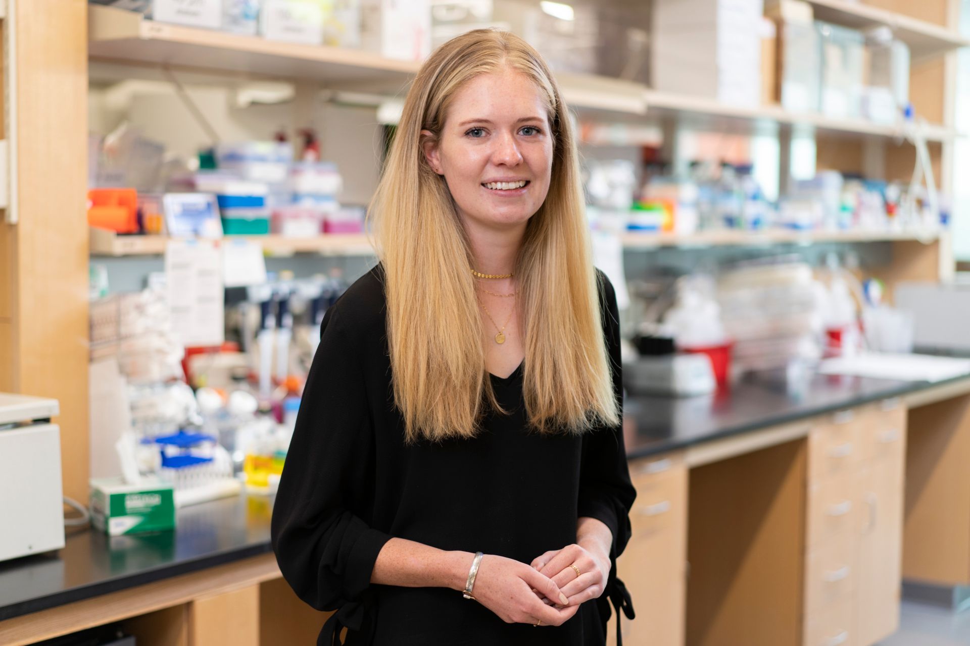
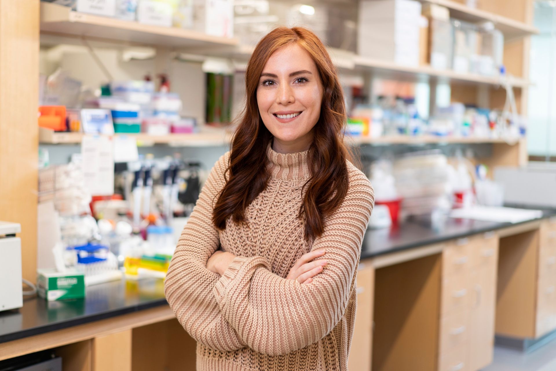
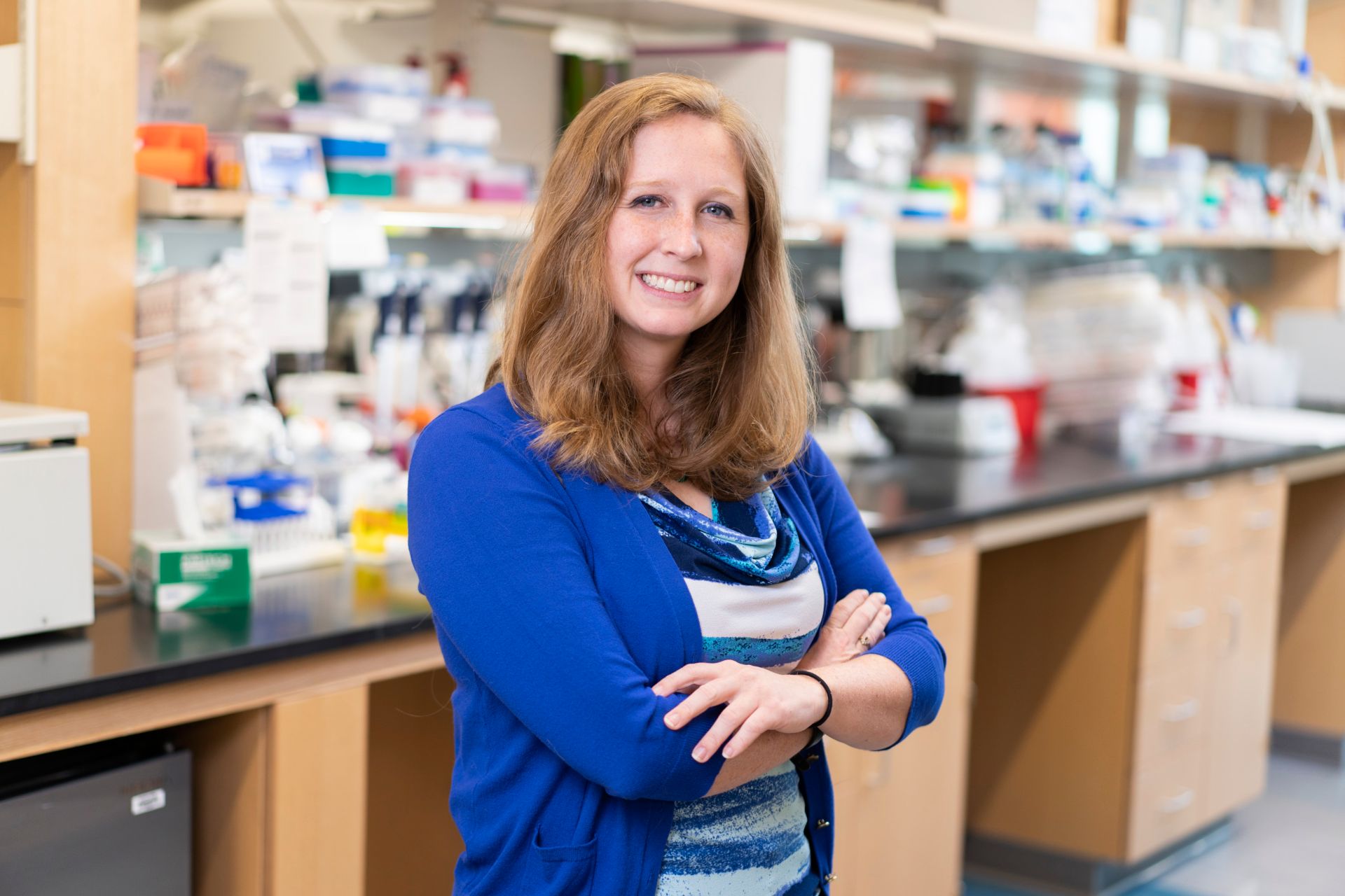
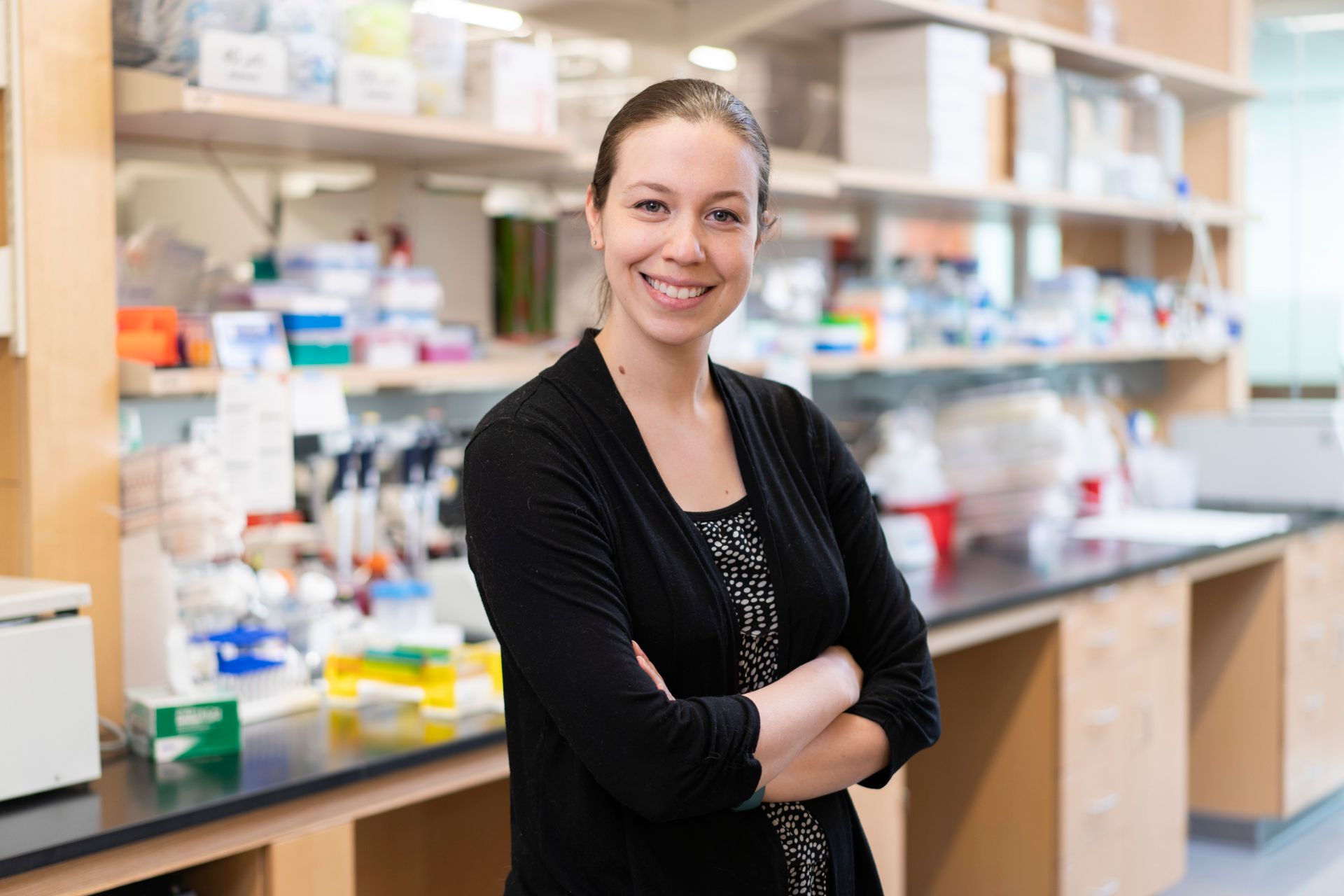
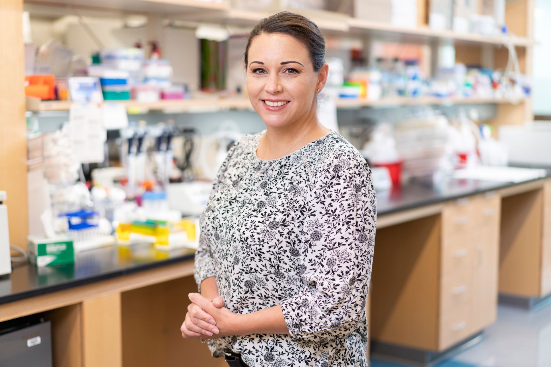
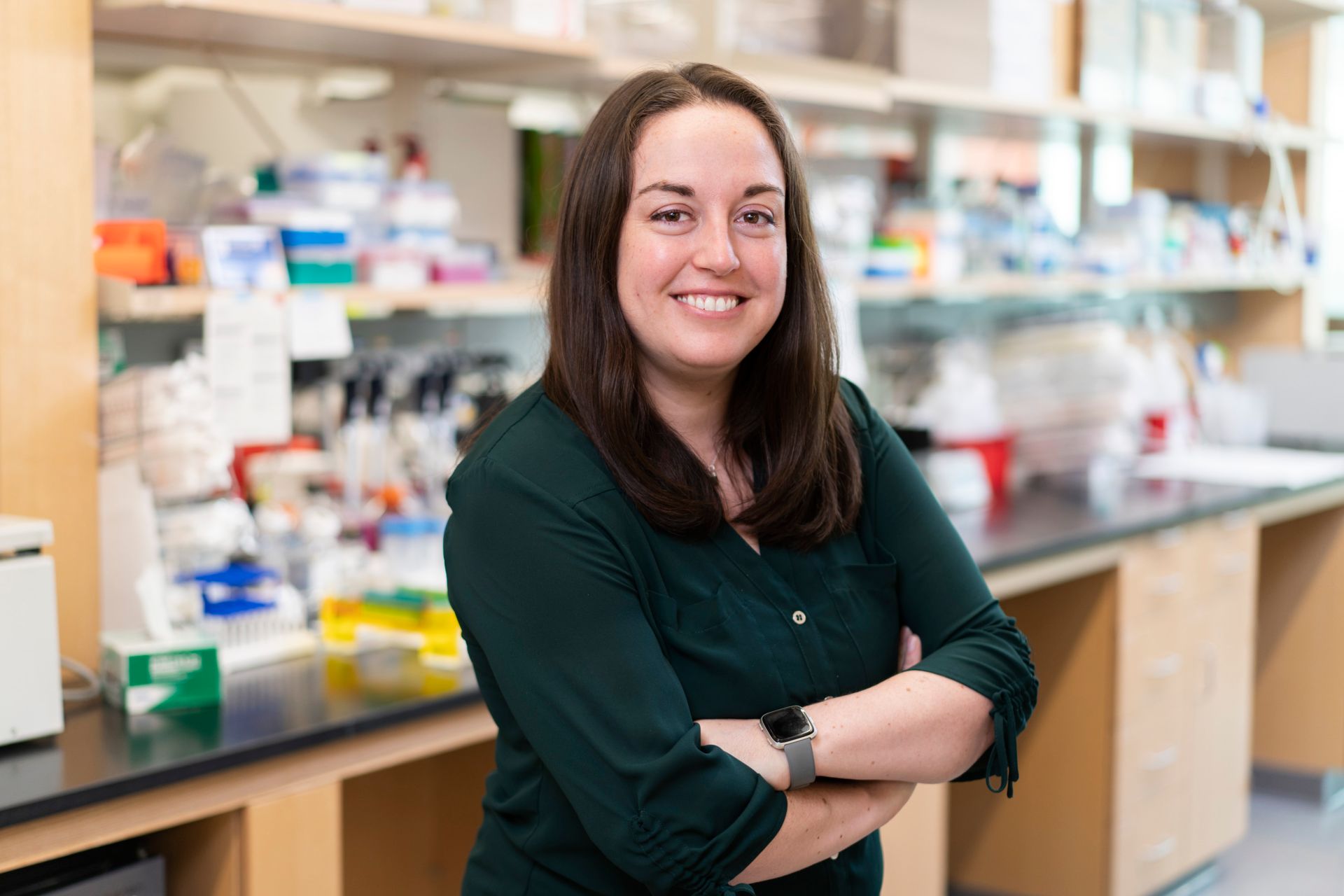
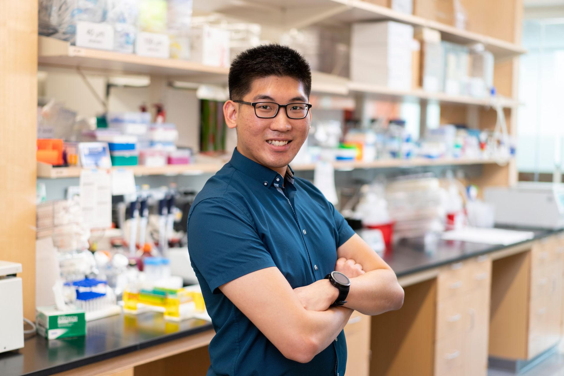
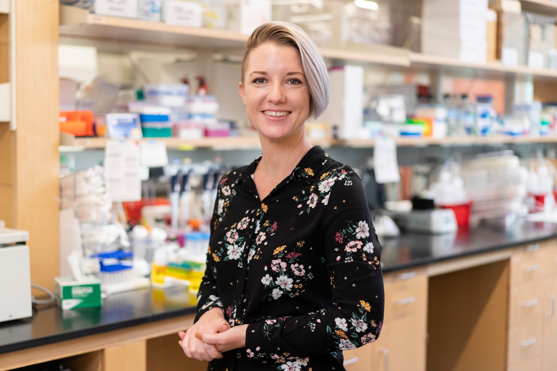
A high-intensity, multidisciplinary team comprised of incredibly diverse individuals that range from research professionals to clinical fellows.



Contact us
Michael Dyer, PhD
Chair & Member
Department of Developmental Neurobiology
MS 324, Room D2039
St. Jude Children's Research Hospital
Follow us

Memphis, TN, 38105-3678 USA GET DIRECTIONS